
Introduction to Venomous snakes and their Physiology
Authored by Kimberly Urban, aka “Dr Viper”
Venomous snakes! Cobra, viper, mamba…these words strike fear into the hearts of many, but fascination and admiration into others. I’ve been working with venomous snakes, primarily vipers, for the past 5 years and other snake species for 10 years. These amazing creatures are a huge part of my life, and I continue to be fascinated by their evolution and adaptability even now. In my series of articles, I will try to introduce you to basic venomous snake biology and physiology, keeper safety and techniques, and delve into husbandry of some of the more common and interesting species.
Venomous snakes are snakes that have evolved modified salivary glands that secrete and store a fluid containing enzymes and toxins. They have modified dentition as well, to allow the delivery of said fluid with a bite. Remember, they are not poisonous!
A poison acts through ingestion, while venom acts through injection. You could very well eat a cobra and not get sick, but if it bites you, you’re in trouble! The physiology of truly poisonous animals, such as frogs and salamanders, is completely different from that of the venomous snakes and lizards.
Not all venomous reptiles are created alike. There are multiple types, or classes, of venom, each acting on specific parts of the body in specific ways. Remember, the purpose of any venom is to immobilize a prey item and begin the digestion process for the snake. Snakes and lizards don’t chew their food, and venomous ones generally do not constrict, so breaking down bones and tougher tissues with venom could potentially enable the animal to digest more efficiently and easily. It is theorized that snakes that have had their venom glands and ducts removed are not able to extract all the nutrients from their food and therefore do not live as long or as healthily as intact specimens. However, venomous reptiles can choose not to inject venom with a bite, or to inject very little, which could challenge this theory! In my collection, I feed mostly frozen-thawed food, and several of my rattlesnakes (Crotalus ssp.) do not even strike, instead choosing to gently accept their meals from my tongs and swallow immediately. I do not think these animals inject much, if any, venom into their food. On the other hand, I have a very large female West African Gaboon viper (Bitis rhinoceros) that injects massive amounts of venom into her frozen-thawed rats, and the rats will bleed even though they’ve been dead for some time!
Hemotoxin
What causes a dead rodent to begin to bleed, even though it has no more circulation? The first class of venom, hemotoxic venom, explains this phenomenon. Hemotoxic venom acts by breaking down red blood cells and vessel walls (hemolytics), disrupting clotting (thrombolytics) and inducing generalized tissue damage (1). Many of the vipers employ hemotoxic venoms, including rattlesnakes, Gaboon vipers, eyelash vipers, and puff adders. A hemotoxic venom is rather slow acting, and often the snake will have to track its prey after injecting venom. The prey item will often suffer hemolytic shock as its blood either begins to flow from pores and orifices (anticoagulants), or coagulates into a gelatin in its veins (procoagulants). Humans bitten by a snake with hemotoxic venom suffer the same fate- disruptions in blood clotting, blister formation (blebbs), blood pressure disturbances, heart rhythm disturbances, and tissue necrosis from the internal bleeding or lack of blood flow
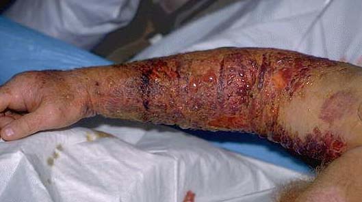
Figure 1. Rattlesnake bite. Courtesy of The Museum of Life and Sciences, http://lifeandscience.org/keepers/2011/02/13/no-such-thing-as-a-poisonous-snake-part-2/.
However, a human often has hours or days to combat the effects of a purely hemotoxic venom before death occurs, and death is often due to infection of the wound area or shock from widespread tissue damage.
Cytotoxin
A cytotoxic venom acts directly on tissues, killing and breaking down cells of the body. Some cytotoxic components include fibrinogenases, collagenases and pshospholipases, which attack and digest cell skeletal proteins, collagen and cell membranes, respectively; cytotoxic venoms literally digest the prey’s tissues
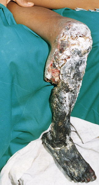
Tissue necrosis resulting from a Bothrops asper bite. Picture courtesy of Gutiérrez JM, Theakston RDG, Warrell DA (2006) Confronting the Neglected Problem of Snake Bite Envenoming: The Need for a Global Partnership. PLoS Med 3(6): e150, doi:10.1371/journal.pmed.0030150
(Figure 2), and are the type of venom thought to aid in digestion in the snakes possessing it (2, 3). Nearly all of the vipers, and some elapids like spitting cobras, monocled cobras, and Indian cobras. Like with the hemotoxic venom, death results from rapid tissue death and organ failure, but may take hours to days in a victim such as a human.
Neurotoxin
A neurotoxic venom, however, results in much more rapid death. With neurotoxic venoms, death occurs from paralysis and respiratory failure, often within minutes. A full-grown human can die from a neurotoxic bite within 15-20 minutes, although most often have up to an hour before full respiratory collapse (3). Neurotoxic venoms can act on the presynaptic neuron to block nerve signals (neurotransmitters) from traveling out of that cell, and these are often much longer-acting and less reversible than postsynaptic neurotoxins, which bind to receptors and keep released neurotransmitters from causing a response in subsequent neurons (4).
It’s All in the Fang
As you can see, not all venoms are created equal; not all venom delivery systems are, either! Snakes have evolved various types of fangs and biting patterns to fit their ecological niches. Opisthoglyphous snakes, such as colubrids, have small, fixed fangs situated in the rear of the mouth, behind the eyes. These are also referred to as rear-fanged snakes (Figure 3B). Opisthoglyph fangs do not have grooves, or only very small, primitive ones, to direct venom, so the snake must “chew” the venom into its prey by biting, holding on, and gnawing (3). This action draws the venom out of the glands and directs it down out of the tooth beds into the prey.
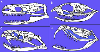
Dendition of snakes. A, aglyphous or non-venomous python skull. B, opisthoglyph, or rear-fanged venomous snake skull. C, proteroglyph, or elapid skull. D, solenoglyph, or viperid skull. Photo courtesy of The Texan Herper: Reptilian II, http://www.thetexanherper.com/2012/07/reptilian-ii.html.
Representative species include boomslangs, twig snakes, vine snakes, and false water cobras. It’s also thought that other aglyphous colubrids, such as garter snakes, are not completely devoid of venom and have small rear fangs; they just are not medically significant to humans (Figure 3A) Proteroglyphs, or elapids, also have small fixed fangs, but their fangs are situated in the front of the mouth, and have large grooves, or are hollow, for directing venom down through the fang and into the prey at the bite site (Figure 3C). While proteroglyphous snakes do not need to chew their venom into prey, they often bite hard and hold on, as their short fangs do not penetrate very deeply into the prey (3).
Solenoglyphs, or vipers, have the most highly evolved venom delivery systems, with large venom glands and massive, hinged fangs. Solenoglyph fangs rest folded against the roof of the mouth when not in use; when the snake strikes, it opens its mouth, and muscles stretch the fangs taut and forward (3)(Figure 3D). A solenoglyph stabs more than it bites, with the fangs often hitting when the mouth is open 180 degrees! That being said, many large vipers are capable of striking with immense force and in snakes such as Gaboon vipers, the trauma of the bite alone can kill small prey. My large female Gaboon viper is 5 feet long and her fangs are easily 2.5 inches long- I shudder to think of the physical trauma a bite from her would cause, not even counting the venom!
While venomous snakes and their powerful arsenal of dentally delivered toxins can seem quite frightening, many components of venom have found medical uses, and are currently saving lives. For example, phospholipase A2 from Cerastes cerastes and Macrovipera lebetina has shown strong anti-tumor activity, and other venom components also show promise for combating cancers (5, 6, 7). Mamba venom neurotoxins are being investigated as pain killers (8). Procoagulants in taipan venom are being used to stop major bleeding during and after surgery (9), and Malayan pit viper (Calloselasma rhodostoma) venom anti-coagulants have shown promise breaking down clots and preventing or treating strokes (10). So you see, snake venoms are vital to the advancement of medicine. Many facilities run “venom lines,” large collections of snakes that are routinely milked for their venom. The venom is then sold or donated to research and medical facilities. This helps to ensure a steady supply of venom to develop not only antivenins, but also to examine components for their utility in treating illnesses and injuries. Hopefully now you understand a bit more about the physiology of venomous reptiles, and that all bites are not created equal!
References
(1). Hemotoxin: proteolytic poison. Online Biology Dictionary. http://www.macroevolution.net/hemotoxin.html.
(2). Bottrall, Joshua L.; Frank Madaras; Christopher D Biven; Michael G Venning; Peter J Mirtschin(2010). Proteolytic activity of Elapid and Viperid Snake venoms and its implication to digestion. Journal of Venom Research 1(3):18–28.PMID 21544178.
(3) (Edited by) Bauchot, Roland (1994). Snakes: A Natural History. New York City, NY, USA: Sterling Publishing Co., Inc. pp. 194–209. ISBN 1-4027-3181-7.
(4) Hodgson, Wayne C.; Wickramaratna, Janith C.(2002). In vitro neuromuscular activity of snake venoms. Clinical and Experimental Pharmacology and Physiology 29 (9): 807–814. PMID 12165047.
(5) Zouari-Kessentini, R; Srairi-Abid, N; Bazaa, A; El Ayeb, M; Luis, J; Marrakchi, N (2013). Antitumoral potential of Tunisian snake venoms secreted phospholipases A2. BioMed Research International: 391389. PMID 23509718
(6) Vyas, V. K.; Brahmbhatt, K; Bhatt, H; Parmar, U; Patidar, R (2013). Therapeutic potential of snake venom in cancer therapy: Current perspectives. Asian Pacific Journal of Tropical Biomedicine 3(2):156–62. PMID 23593597
(7) Jain, D; Kumar, S (2012). Snake venom: A potent anticancer agent. Asian Pacific journal of cancer prevention : APJCP 13 (10): 4855–60. PMID 23244070
(8) Craik DJ, Schroeder CI (2013). Peptides from mamba venom as pain killers. Angew Chem Int Ed Engl; 52(11):3071-3. Epub 2013 Feb 4. PMID 23382094
(9) Marsh, NA (2001). Diagnostic uses of snake venom. Haemostasis; 31(3-6):211-7. PMID 11910187
(10) Jahnke H (1991). Experimental ancrod (Arvin) for acute ischemic stroke: nursing implications. J Neurosci Nurs; 23(6):386-9. Review. PMID 1839548